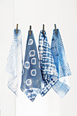
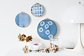
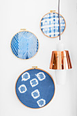
12322297 - Old handkerchiefs hand-dyed using Shibori technique hung from line12322294 - Hand-made wall decorations made from embroidery rings and old handkerchiefs hand-dyed using Shibori technique71335930 - Itsukushima Miyajima Japan. Handkerchiefs12322295 - Hand-made wall decorations made from embroidery rings and old handkerchiefs hand-dyed using Shibori technique13584120 - Stitched handkerchiefs at window with table set for Easter13354555 - Baked 'handkerchiefs' with flowers and walnuts10251947 - Bed with patchwork quilt in simple bedroom11326213 - Posy of forget.me-not wrapped in white cloth12322291 - Gift wrapping made from old handkerchief and hand-dyed using Shibori technique10185661 - Portrait of gray eyed Charlotte woman with blonde hair cleaning her face, smiling12424687 - Preparations for dying an old handkerchief using the Shibori technique: fold fabric in concertina and fix with clothes pegs11326226 - Cut out paper hand and forget-me-not soap on cloth with forget-me-not motif13780084 - Paper Flowers11128188 - Fine embroidered white cotton hankerchiefs hung from wooden pegs11326225 - Forget-me-not soap and cut out paper parasol on cloth with forget-me-not motif13995237 - Woman Blotting Lipstick13718702 - Boy sneezing13799165 - Middle-aged African-American man with headache taking his medicine13726198 - Mother applying medicine to sick boy13995632 - Variety of medications on bedside table,studio shot13894270 - Portrait of Man Celebrating His Birthday11169836 - A woman with a cold drinking tea in bed13894271 - Man Shaking Birthday Present13995633 - Medication and crumpled facial tissues and alarm clock on bedside table,studio shot13995634 - Medications,bowl of chicken soup and glass of water on table,studio shot13921848 - Young woman lying on stomach on the grass sneezing\n13781086 - Man Blowing Nose13799164 - Middle-aged African-American man with headache taking his medicine00718796 - Peonies in a vase on a lace cloth13726199 - Mother applying medicine to sick boy13925092 - The image presents oak xylem tissue in the transversal cross-section of the stalk, photographed through the microscope in polarized light at a magnification of 400X\n00946481 - Young woman blowing her nose13924655 - The image presents stomata in lily leaf epidermis, photographed through the microscope in polarized light at a magnification of 200X\n13799161 - Close-up shot of senior African-American man with a cold blowing his nose13726203 - Sick boy lying in bed13925405 - The image presents stomata in Spathiphyllum sp. leaf epidermis, photographed through the microscope in polarized light at a magnification of 100X\n13925048 - The image presents stomata in Croton leaf epidermis, photographed through the microscope in polarized light at a magnification of 100X\n13924924 - The image presents stomata in Croton leaf epidermis, photographed through the microscope in polarized light at a magnification of 200X\n13924686 - The image presents nettle tissues in longitudinal cross-section of the stalk, photographed through the microscope in polarized light at a magnification of 100X. Small, round orange particles are the crystals of calcium oxalate called druses.\n13925332 - The image presents stomata in hosta leaf epidermis, photographed through the microscope in polarized light at a magnification of 100X\n13799162 - Close-up shot of senior African-American man with a cold blowing his nose13925454 - The image presents nettle tissues in the stalk in longitudinal cross-section, photographed through the microscope at a magnification of 100X\n13925345 - The image presents nettle tissues in the stalk in transversall cross-section, photographed through the microscope in polarized light and dark field at a magnification of 100X.\n13925287 - The image presents tissues in nettle stalk in longitudinal cross-section, photographed through the microscope in polarized light at a magnification of 100X\n13925093 - The image presents a single vascular bundle in Carex sp. stalk, photographed through the microscope in polarized light and dark field at a magnification of 200X\n13925038 - The image presents stomata in Spathiphyllum leaf epidermis, photographed through the microscope in polarized light at a magnification of 200X\n13718703 - Boy sneezing13924302 - The image presents nettle tissues in the transversal section of the stalk, photographed through the microscope in polarized light at a magnification of 400X\n13925240 - The image presents stomata in Spathiphyllum leaf epidermis, photographed through the microscope in polarized light at a magnification of 200X\n13925070 - The image presents stomata in Spathiphyllum sp. leaf epidermis, photographed through the microscope in polarized light at a magnification of 100X\n13925303 - The image presents stomata in Spathiphyllum leaf epidermis, photographed through the microscope in polarized light at a magnification of 400X\n11080642 - Clothes pegs decorated with tape on metal number stencil next to decorative vintage bottles13925579 - The image presents stomata in Spathiphyllum leaf epidermis, photographed through the microscope in polarized light at a magnification of 200X\n13925085 - The image presents vascular bundles in senecio stalk, photographed through the microscope in polarized light at a magnification of 200X\n13925078 - The image presents stomata in lily leaf epidermis, photographed through the microscope in polarized light at a magnification of 200X\n13925010 - The image presents read leaf in transversal cross-section, photographed through the microscope in polarized light at a magnification of 100X\n13924841 - The image presents tissues in nettle stalk in longitudinal cross-section, photographed through the microscope in polarized light at a magnification of 100X. The round yellow structures on the bottom are druses. Druses are the structures created by calcium oxalat.\n13726196 - Sick boy lying in bed13925265 - The image presents nettle tissues in the stalk in longitudinal cross-section, photographed through the microscope in polarized light at a magnification of 100X\n11053751 - Dried poppy seed heads between glass containers of cotton wool and tissues13925259 - The image presents stomata in hyacinth leaf epidermis, photographed through the microscope in polarized light at a magnification of 100X\n13925106 - The image presents reed stalk in the transversal cross-section, photographed through the microscope in polarized light and dark field, at a magnification of 100X\n13925468 - The image presents palisade mesophyll in hyacinthus leaf (transversal cross-section) photographed through the microscope in polarized light at a magnification of 200X\n13925616 - The image presents stomata in Stromanthe sp. leaf epidermis, photographed through the microscope in polarized light at a magnification of 100X\n13572751 - Small gift wrapped in handkerchief11163341 - Woman blowing her nose13925009 - The image presents nettle stalk longitudinal cross-section photographed through the microscope in polarized light at a magnification of 100X\n13924555 - The image presents tissues in nettle stalk in longitudinal cross-section, photographed through the microscope in polarized light at a magnification of 100X\n13924337 - The image presents tissues in nettle stalk in longitudinal cross section, photographed through the microscope in polarized light at a magnification of 100X\n13925580 - The image presents Anemone sylvestris stalk in transversal cross-section, photographed through the microscope in polarized light at a magnification of 200X\n13925297 - The image presents stomata in Spathiphyllum leaf epidermis, photographed through the microscope in polarized light at a magnification of 100X\n13924900 - The image presents stomata in Spathiphyllum sp. leaf epidermis, photographed through the microscope in polarized light at a magnification of 100X\n13924564 - The image presents stomata in Spathiphyllum leaf epidermis, photographed through the microscope in polarized light at a magnification of 200X\n13925148 - The image presents stomata in Spathiphyllum leaf epidermis, photographed through the microscope in polarized light at a magnification of 400X\n13924866 - The image presents reed stalk in transversal cross-section, photographed through the microscope in polarized light at a magnification of 200X\n13924539 - The image presents stomata in Spathiphyllum leaf epidermis, photographed through the microscope in polarized light at a magnification of 200X\n13924552 - The image presents stomata in Spathiphyllum leaf epidermis, photographed through the microscope in polarized light at a magnification of 400X\n13924490 - The image presents tissues in nettle stalk in longitudinal cross-section, photographed through the microscope in polarized light at a magnification of 100X. The round yellow structures are druses. Druses are the structures created by calcium oxalate.\n13925464 - The image presents stomata in Stromanthe sp. leaf epidermis, photographed through the microscope in polarized light at a magnification of 100X\n13925083 - The image presents Utricularia trap, a kind of carnivorous plant, photographed through the microscope in polarized light and dark field, at a magnification of 100X\n13924698 - The image presents nettle tissues in the stalk in longitudinal cross-section, photographed through the microscope at a magnification of 100X\n00936528 - Young woman blowing her nose13924921 - The image presents senecio tissues in the transversal cross-section through the stalk, photographed through the microscope in polarized light and dark field at a magnification of 100X\n13924624 - The image presents a single stoma in Spathiphyllum sp. leaf epidermis, photographed through the microscope in polarized light at a magnification of 200X\n13925525 - The image presents Carex sp. leaf in transversal cross-section, photographed through the microscope in polarized light at a magnification of 100X\n13925241 - The image presents nettle tisues in the transversal cross-section of the stalk, photographed through the microscope in polarized light at a magnification of 200X\n13924796 - The image presents knautia arvensis tissues in the transversal section of the stalk, photographed through the microscope in bright field, at a magnification of 100X\n13924748 - The image presents a single stoma in Knautia arvensis epidermis, photographed through the microscope in polarized light at a magnification of 200X\n13924580 - The image presents reed stalk in transversal cross-section, photographed through the microscope in bright field, at a magnification of 100X\n13925392 - The image presents reed stalk in transversal cross-section, photographed through the microscope in polarized light at a magnification of 200X\n13925207 - The image presents stomata in Spathiphyllum leaf epidermis, photographed through the microscope in polarized light at a magnification of 400X\n13924809 - The image presents nettle tissues in the transversal cross-section of the stalk, photographed through the microscope in polarized light at a magnification of 200X\n13924786 - The image presents knautia arvensis tissues in the transversal section of the stalk, photographed through the microscope in bright field, at a magnification of 100X\n13925549 - The image presents a small part of forsythia stalk tissues in transversal cross-section, photographed through the microscope in polarized light at a magnification of 100X\n13925473 - The image presents stomata in Spathiphyllum leaf epidermis, photographed through the microscope in polarized light at a magnification of 100X\n13924392 - Tissues in nettle stalk photographed through the microscope\n13924913 - The image presents stomata in Spathiphyllum leaf epidermis, photographed through the microscope in polarized light at a magnification of 200X\n13924452 - The image presents reed stalk in transversal cross-section, photographed through the microscope in polarized light at a magnification of 200X\n13925603 - The image presents oak tissues in transversal cross-section of the stalk, photographed through the microscope in polarized light at a magnification of 100X\nnext page