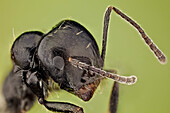
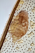
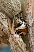
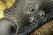
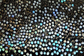
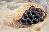
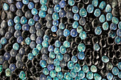
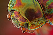
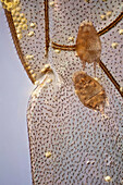
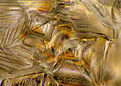
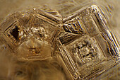
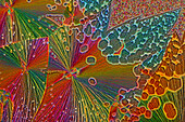
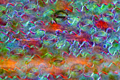
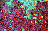
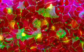
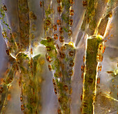
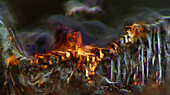
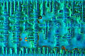
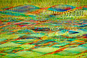
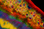
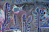
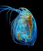
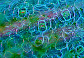
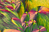
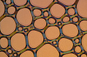
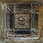
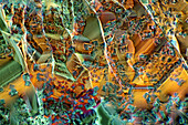
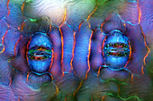
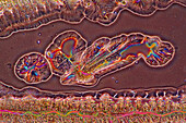
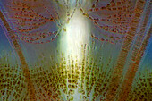
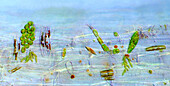
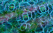
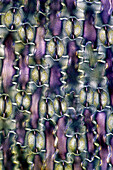
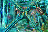
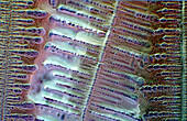
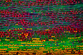
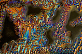
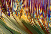
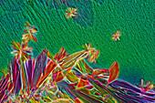
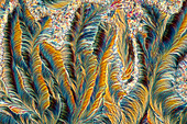
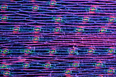
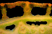
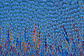
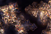
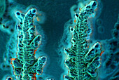
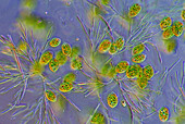
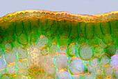
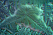
71103624 - 'A high magnification picture of the ocelli and ommatidium of a german wasp; a special technique to get a SEM like image was used.'13847357 - High magnification shot of an ant, it shows how hairy they can be13848992 - A detail of a Parasitic mite on a bee wing, it also show the texture of some pollen grains; 100:1 magnification13844800 - Detail of the bug nymph at higher magnification, holding itself on a dried wild flower13845189 - A high magnification picture of the ocelli and ommatidium of a german wasp; a special technique to get a SEM like image was used.13845657 - These weevils are among the most colorfull ones in the world; high magnification detail of the scales in the thorax13849299 - Very high magnification shot of a silverfish eye; a special technique was used to reveal all the eye surface texture in order to get a SEM like image, but with colour.13844865 - These weevils are among the most colorfull ones in the world, very high magnification shot of the scales. They do look like jewels13846973 - A head view of Chrysolina americana; magnification 6:113845015 - A bee wing covered on pollen showing two parasitic mites; 50:1 magnification13925413 - The image presents recrystallized salt, photographed through the microscope in polarized light at a magnification of 100X\n13925092 - The image presents oak xylem tissue in the transversal cross-section of the stalk, photographed through the microscope in polarized light at a magnification of 400X\n13924986 - The image presents crystallized tartaric acid, photographed through the microscope in polarized light at a magnification of 100X\n13924422 - The image presents crystallized mixture of kitchen salt and erythritol, photographed through the microscope in polarized light at a magnification of 100X\n13924309 - The image presents crystallized soy sauce photographed through the microscope in bright field at a magnification of 100X\n13925446 - The image presents crystals of recrystallized kitchen salt, photographed through the microscope in polarized light at a magnification of 100X\n13925098 - The image presents crystallized mixture of urea and paracetamol, photographed through the microscope in polarized light at a magnification of 100X\n13924754 - The image presents crystallized mixture of malic acid and hydroquinone photographed through the microscope in polarized light at a magnification of 100X\n13924655 - The image presents stomata in lily leaf epidermis, photographed through the microscope in polarized light at a magnification of 200X\n13924442 - The image presents tiny air bubbles photographed through the microscope in polarized light at a magnification of 100X\n13925480 - The image presents crystallized mixture of urea and malic acid, photographed through the microscope in polarized light at a magnification of 100X\n13925405 - The image presents stomata in Spathiphyllum sp. leaf epidermis, photographed through the microscope in polarized light at a magnification of 100X\n13925069 - The image presents diptera larva, photographed through the microscope in polarized light at a magnification of 100X\n13925048 - The image presents stomata in Croton leaf epidermis, photographed through the microscope in polarized light at a magnification of 100X\n13924963 - The image presents crystallized resorcinol, photographed through the microscope in polarized light at a magnification of 100X\n13924924 - The image presents stomata in Croton leaf epidermis, photographed through the microscope in polarized light at a magnification of 200X\n13924859 - "The image presents Cladophora sp. ""twigs"" (a kind of green algae) with Cocconeis sp. (a kin of diatoms) settled on it, photographed through the microscope in polarized light at a magnification of 200X"\n13924764 - The image presents Batrachospermum sp., a kind of red algae, photographed through the microscope in polarized light at a magnification of 200X\n13924686 - The image presents nettle tissues in longitudinal cross-section of the stalk, photographed through the microscope in polarized light at a magnification of 100X. Small, round orange particles are the crystals of calcium oxalate called druses.\n13924465 - The image presents crystallized silver nitrate, photographed through the microscope in polarized light at a magnification of 100X\n13924398 - The image presents recrystallized sugar photographed through the microscope in polarized light at a magnification of 100X\n13924389 - The image presents recrystallized mixture of salt and erithrytol, photographed through the microscope in polarized light at a magnification of 100X\n13925423 - The image presentstwo suctorians ( a kind of ciliate) and tiny diatoms, photographed through the microscope in polarized light at a magnification of 200X\n13925385 - The image presents Fragilaria sp., a kind of diatoms against Batrachospermum, a kind of red algae, photographed through the microscope in polarized light at a magnification of 200X\n13925332 - The image presents stomata in hosta leaf epidermis, photographed through the microscope in polarized light at a magnification of 100X\n13925277 - The image presents crystallized sulfur and hydroquinone, photographed through the microscope in polarized light at a magnification of 100X\n13924981 - The image presents crystallized mixture of kitchen salt and erythritol, photographed through the microscope in polarized light at a magnification of 100X\n13924950 - The image presents crystallized soy sauce, photographed through the microscope in polarized light at a magnification of 100X\n13924827 - The image presents crystallized soy sauce, photographed through the microscope in polarized light at a magnification of 100X\n13925454 - The image presents nettle tissues in the stalk in longitudinal cross-section, photographed through the microscope at a magnification of 100X\n13925345 - The image presents nettle tissues in the stalk in transversall cross-section, photographed through the microscope in polarized light and dark field at a magnification of 100X.\n13925287 - The image presents tissues in nettle stalk in longitudinal cross-section, photographed through the microscope in polarized light at a magnification of 100X\n13925093 - The image presents a single vascular bundle in Carex sp. stalk, photographed through the microscope in polarized light and dark field at a magnification of 200X\n13925044 - The image presents various tiny algae settled on Lemna sp. root, photographed through the microscope in polarized light at a magnification of 200X\n13925038 - The image presents stomata in Spathiphyllum leaf epidermis, photographed through the microscope in polarized light at a magnification of 200X\n13924496 - The image presents crystallized soy sauce, photographed through the microscope in polarized light at a magnification of 100X\n13925404 - The image presents crystallized soy sauce, photographed through the microscope in polarized light at a magnification of 100X\n13925205 - The image presents crystallized soy sauce, photographed through the microscope in polarized light at a magnification of 100X\n13925162 - The image presents crystallized soy sauce, photographed through the microscope in polarized light at a magnification of 100X\n13924884 - The image presents a single crystal of recrystallized kitchen salt, photographed through the microscope in polarized light at a magnification of 200X\n13924619 - The image presents crystallized tartaric acid, photographed through the microscope in polarized light at a magnification of 100X\n13924612 - The image presents crystals of recrystallized kitchen salt, photographed through the microscope in polarized light at a magnification of 100X\n13924302 - The image presents nettle tissues in the transversal section of the stalk, photographed through the microscope in polarized light at a magnification of 400X\n13925240 - The image presents stomata in Spathiphyllum leaf epidermis, photographed through the microscope in polarized light at a magnification of 200X\n13925169 - The image presents crystallized soy sauce photographed through the microscope in polarized light at a magnification of 100X\n13925163 - The image presents Simocephalus sp., a kind of cladoceran, photographed through the microscope in polarized light at a magnification of 100X\n13925161 - The image presents red wine photographed through the microscope in polarized light at a magnification of 100X\n13925070 - The image presents stomata in Spathiphyllum sp. leaf epidermis, photographed through the microscope in polarized light at a magnification of 100X\n13924994 - The image presents crystallized mixture of erythritol and paracetamol, photographed through the microscope in polarized light at a magnification of 100X\n13924573 - The image presents air bubbles formed in foemaed milk photographed through the microscope in polarized light at a magnification of 100X\n13924411 - The image presents a single crystal of recrystallized salt, photographed through the microscope in polarized light at a magnification of 100X\n13924379 - The image presents mixture of sugar and salt, crystallized photographed through the microscope in polarized light at a magnification of 100X\n13925303 - The image presents stomata in Spathiphyllum leaf epidermis, photographed through the microscope in polarized light at a magnification of 400X\n13924965 - The image presents crystallized copper acetate photographed through the microscope in polarized light at a magnification of 100X\n13924803 - The image presents Fragilaria sp., a kind of diatoms against Batrachospermum, a kind of red algae, photographed through the microscope in polarized light at a magnification of 200X\n13924513 - The image presents various tiny algae settled on Lemna sp. root, photographed through the microscope in polarized light at a magnification of 200X\n13925579 - The image presents stomata in Spathiphyllum leaf epidermis, photographed through the microscope in polarized light at a magnification of 200X\n13925384 - The image presents air bubbles photographed through the microscope in polarized light at a magnification of 100X\n13925278 - The image presents various tiny algae settled on Lemna sp. root, photographed through the microscope in polarized light at a magnification of 400X. On the right are visible diatoms closed in a special protecting case.\n13925233 - The image presents crystallized resorcinol photographed through the microscope in polarized light at a magnification of 100X\n13925095 - The image presents crystallized soy sauce, photographed through the microscope in polarized light at a magnification of 100X\n13925085 - The image presents vascular bundles in senecio stalk, photographed through the microscope in polarized light at a magnification of 200X\n13925078 - The image presents stomata in lily leaf epidermis, photographed through the microscope in polarized light at a magnification of 200X\n13925010 - The image presents read leaf in transversal cross-section, photographed through the microscope in polarized light at a magnification of 100X\n13924964 - The image presents crystallized mixture of kitchen salt and erythritol, photographed through the microscope in polarized light at a magnification of 100X\n13924896 - The image presents crystallized soy sauce, photographed through the microscope in polarized light at a magnification of 100X\n13924841 - The image presents tissues in nettle stalk in longitudinal cross-section, photographed through the microscope in polarized light at a magnification of 100X. The round yellow structures on the bottom are druses. Druses are the structures created by calcium oxalat.\n13924602 - The image presents Fragilaria sp., a kind of diatoms and Batrachospermum sp., a kind of red algae, photographed through the microscope in polarized light at a magnification of 200X\n13924505 - The image presents crystallized soy sauce, photographed through the microscope in polarized light at a magnification of 100X\n13924417 - The image presents crystallized soy sauce, photographed through the microscope in polarized light at a magnification of 100X\n13925265 - The image presents nettle tissues in the stalk in longitudinal cross-section, photographed through the microscope in polarized light at a magnification of 100X\n13925042 - The image presents crystallized mixture of sugar and salt, photographed through the microscope in polarized light at a magnification of 100X\n13924998 - The image presents crystallized resorcinol, photographed through the microscope in polarized light at a magnification of 100X\n13924640 - The image presents crystallized mixture of myoinositol and tartaric acid, photographed through the microscope in polarized light at a magnification of 100X\n13924355 - The image presents crystallized mixture of paracetamol and resorcinol, photographed through the microscope in polarized light at a magnification of 100X\n13925313 - The image presents crystallized paracetamol, photographed through the microscope in polarized light at a magnification of 100X\n13925259 - The image presents stomata in hyacinth leaf epidermis, photographed through the microscope in polarized light at a magnification of 100X\n13925106 - The image presents reed stalk in the transversal cross-section, photographed through the microscope in polarized light and dark field, at a magnification of 100X\n13924829 - The image presents crystallized mixture of erythritol and TRIS, photographed through the microscope in polarized light at a magnification of 100X\n13924768 - The image presents crystallized mixture os sugar and salt, photographed through the microscope in polarized light at a magnification of 100X\n13924733 - The image presents crystallized soy sauce, photographed through the microscope in bright field at a magnification of 100X\n13924592 - The image presents crystallized soy sauce, photographed through the microscope in polarized light at a magnification of 100X\n13924404 - The image presents euglenoids among green algae, photographed through the microscope in polarized light at a magnification of 100X\n13925468 - The image presents palisade mesophyll in hyacinthus leaf (transversal cross-section) photographed through the microscope in polarized light at a magnification of 200X\n13925453 - The image presents crystallized soy sauce, photographed through the microscope in polarized light at a magnification of 100X\n13925372 - The image presents a single crystal of recrystallized kitchen salt, photographed through the microscope in polarized light at a magnification of 200X\n13925035 - The image presents crystallized mixture of kitchen salt and erythritol, photographed through the microscope in polarized light at a magnification of 100X\n13924918 - The image presents crystallized soy sauce, photographed through the microscope in polarized light at a magnification of 100X\n13924882 - The image presents crystallized soy sauce, photographed through the microscope in polarized light at a magnification of 100X\nnext page